


A lung nodule is described as a solid formation, almost reminiscent of a marble, which affixes itself to the tissue in the lung. The tissue comprises numerous blood vessels and minuscule air sacs in a spongy consistency that supplies the body with oxygen.
While a nodule typically remains symptom-free, there is a possibility it can throw a wrench in the lung’s operational process depending on its size. Also, based on that size will determine whether the formation is cancerous. The procedures when a patient is found to have one of these growths within their cavity is as follows:
- Typically, those that measure below a diameter of 6 mm will involve follow-up CT scans to monitor the growth process since the likelihood for cancer at this size is less than 10% unless the doctor finds an indication that would lead to a higher probability.
- Any nodules above 6 mm but less than 10 mm will be assessed carefully. These are growing close to the requirement for biopsy, which is over 10 mm.
- When a nodule rises above 10 mm, the physician should either remove it or, at the very least, biopsy since these carry an 80% probability for malignancy.
- Anything at or above 3 cm references as a “lung mass.”
Many things can lead to the formation of nodules such as scars or cysts, an impacted airway, old infection or pneumonia, lymph nodes that become enlarged, and cancer can produce these formations.
Diagnosing A Lung Nodule
As a rule, a lung nodule won’t produce any symptoms meaning you can have a formation and be completely unaware. Generally, these are caught when you have an annual physical or another medical procedure where the growth is inadvertently captured from diagnostic testing on another region of the body.
The doctor will then do a specific chest x-ray or CT scan to get the specifics on the nodule so a care plan can be determined. Usually, the chest x-ray is the first step, but the CT scan is performed for a more thorough analysis if the x-ray is not adequate.
The surgeon will go over the results to determine whether biopsy or removal is preferred.
The size is the designating factor. Some diagnostic tools like Tool-in-lesion are innovative, while others remain conventional. Depending on the doctor’s foray into new practices will designate which will be incorporated. Let’s look at a few procedures implemented for nodules today.
● Chest x-ray
With a chest x-ray, invisible radiation produces energy beams to capture images of the lungs to determine if there are formations present. Monitoring of growth occurs with repeat x-rays and CT scans to ensure there is no rapid growth beyond the limits for malignancy.
If it remains consistent for as long as five years, the surgeon can declare it benign growth. The surgeon will suggest a biopsy when there is growth or family history, or if you smoke or perhaps have symptoms.
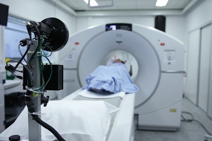
● Chest CT Scan
These scans combine computer technology with x-ray to give a more detailed approach for images of the muscle, organs, bones, and fat. The imagery is often used as a guide to help surgeons when attempting to place needles in the case of a biopsy.
● A biopsy
A closed biopsy occurs via skin insertion or through the windpipe. The open option takes place under general anesthesia in a surgical facility.
● Needle biopsy
A surgeon will use a hollow, thin needle to insert the nodule while viewing the lungs on a CT scan. A sample of the growth is taken for analysis under a microscope.
These also reference as “percutaneous” biopsies meaning they go through the skin or “closed transthoracic.” The doctor will be limited in their capabilities depending on the size of the nodule. If these are tiny, it can be difficult or if the location is challenging to reach.
● Navigational Bronchoscopy
This relatively innovative tool is meant to reach deeper into the cavity and capture the minutest formations. The method is described as (quote) “a GPS-style guided technique combined with a bronchoscope.” Click here to learn about a bronchoscopy.
● Thoracoscopic biopsy
The procedure references a (VATS) or “video-assisted thoracic surgical biopsy. The surgeon will make a small incision in the patient’s chest wall where a video camera attached to a tube inserts.
The process requires staying in the hospital since it is a surgical procedure due to the incision and a portion of the lung being removed.
The surgeon has the capacity to view the inner ribs as well as the outermost part of the lungs while manipulating the camera allowing the surgeon to remove abnormalities for later viewing under the microscope.
Final Thought
Depending on its size, a lung nodule is not something that you will typically find due to symptomology or a visit to the doctor.
Generally, these are inadvertently caught when having either an annual physical or diagnostic testing on another body region. Go to https://www.medicalnewstoday.com/articles/317874/ for guidance on various lung biopsies to learn why you have a nodule.
The primary consideration is the size of the formation. The smaller, the better. If it remains below 6 mm for a period of five years or more, the surgeon will likely classify the growth as benign.
However, suppose it steadily grows as the doctor repeatedly monitors its progression. In that case, there will be a decision to either, go ahead and remove it to eliminate the threat of malignancy or biopsy to determine the same.
Anything over 10 mm has an 80% likelihood for malignancy. Surgical removal makes sense in these instances over biopsy.


